OU students share eye research at 23rd annual SUPER Symposium
 August 08, 2025
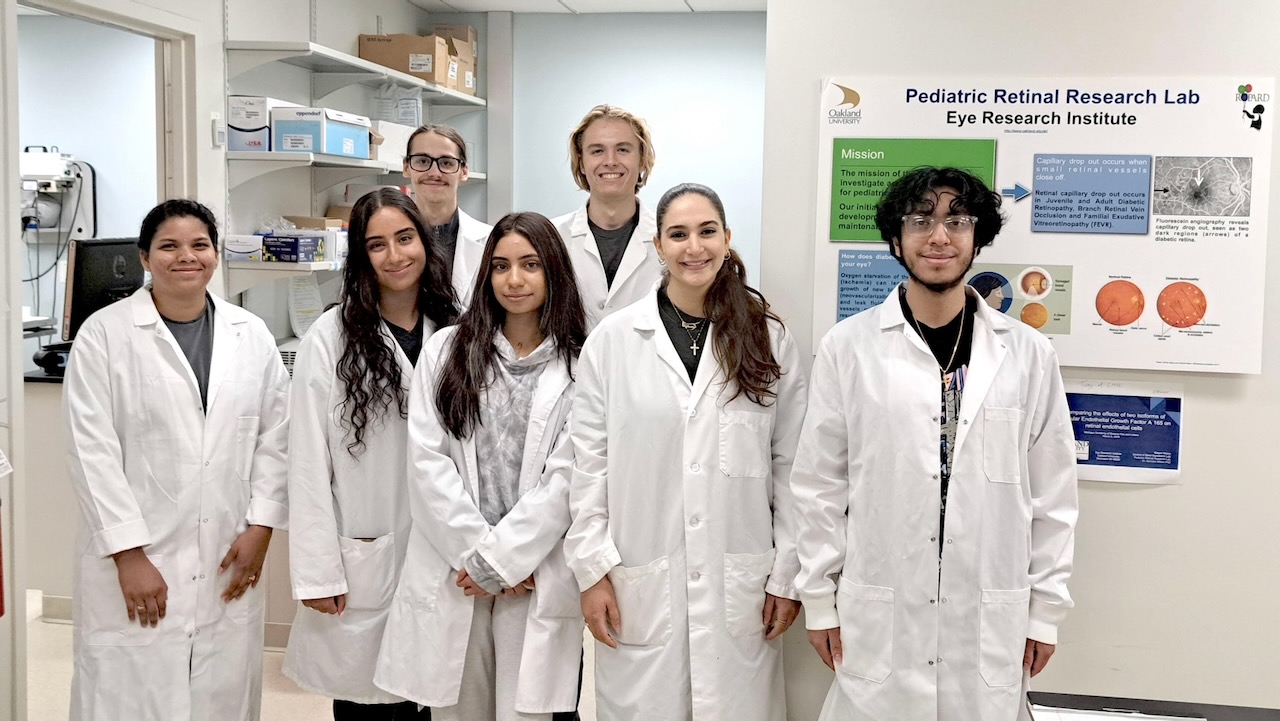
The Summer Undergraduate Program in Eye Research (SUPER) at Oakland University held its 23rd annual student research symposium on Wednesday, July 30 in the Oakland Center.
The highly selective 12-week program allows students to perform research on the causes and potential cures for eyes diseases with guidance from faculty members in the university’s Eye Research Institute (ERI).
“While many university summer undergraduate training programs have been cut down to 10 weeks duration from 12, our sponsor support so far has allowed us to maintain the traditional 12 week program for OU SUPER students,” said Dr. Kenneth Mitton, associate professor of biomedical sciences (ERI) and director of the SUPER program.
Using the latest scientific methodologies and equipment, the students spent at least 30 hours per week conducting lab experiments and attending a weekly Vision Science seminar given by ERI and Beaumont Ophthalmology faculty. The students also received a research fellowship, as well as training in professional speaking and presentation.
This year’s SUPER students include:
• Alex Sanchez, a junior who is pursuing a degree in biochemistry. Sanchez was mentored by Dr. Michael Risner.
“The basics of my research was that I wanted to see if the protein Equinatoxin II binds to sphingomyelin, which is a lipid.” Sanchez said. “I produced the Equinatoxin II, confirmed that what I produced was Equinatoxin II, and performed tests to see if Equinatoxin II is actually binding to sphingomyelin only.”
• Sudharshini Nayudu, a junior who is pursuing a degree in biomedical sciences. Nayudo was mentored by Dr. Michael Risner.
“This summer at Oakland University’s Eye Research Institute, I studied how the enzyme acid sphingomyelinase (ASM) affects retinal function using electroretinography (ERG),” Nayudo said. “ASM is important in lipid metabolism and inflammation, and we focused on its role in retinal ganglion cells (RGCs), which are involved in glaucoma.
“Using genetically modified mice with different ASM levels in Thy1+ retinal cells, we analyzed ERG waveforms to see how ASM impacts retinal responses to light,” Nayudu added. “Our results suggest that ASM activity may contribute to RGC degeneration, making it a potential target for glaucoma treatment.”
• Michael Binkowski, a junior who is majoring in clinical and diagnostic science in medical laboratory science. Binkowski was mentored by Dr. Mohamed Al-Shabrawey.
“Diabetic Retinopathy (DR) is primarily caused by/ worsened by permeable blood vessels causing their fluid to leak onto the retina,” Binkowski said. “Current therapies for DR are often invasive, costly, etc. A newly proposed treatment for DR is inhibition of the Renin-Angiotensin system. While RAS inhibition can prevent the onset of DR, it does not prevent the progression of it.
“We showed that if certain aspects of the RAS were inhibited, like in the proposed treatments, there could be an increase in EETs, which increase the permeability of retinal blood vessels, which is not good for DR. This increase in EETs may explain why RAS inhibition may not prevent the progression of DR.”
• Gabriella Kirakos, a sophomore who is majoring in clinical and diagnostic sciences in pre-clinical professions. Kirakos was mentored by Dr. Dao-Qi Zhang.
“This summer, I worked in Dr. Zhang’s lab at the Eye Research Institute, where I studied the structure and function of the retina using molecular, morphological, and functional techniques,” Kirakos said. “My project involved three main methods: Genotyping to identify transgenic mice with fluorescent tags in specific retinal cells, Immunohistochemistry to label and visualize those cells in retina tissue slices using antibodies, and Electroretinography to measure how well different retinal cells respond to light. By combining these methods, researchers can better understand how different retinal cells develop, are organized, and work together to support vision.”
• Johnathan Stark, a senior who is pursuing a degree in physics. Stark was mentored by Dr. Kenneth Mitton.
“At the SUPER 2025 symposium, I presented research on the prevalence of diseases that affect the blood vessels in the retina such as, diabetic retinopathy and retinopathy of prematurity, as well as preclinical research of Noregen,” Stark said. “The Mitton lab, alongside Dr. Kimberly Drenser, has been researching the effects of a protein called Norrin which plays a key role in helping the blood vessels of the retina grow properly. The Mitton lab, with Retinova Therapeutics, developed Noregen, a version of Norrin that has been purified to make it safe for use in living systems. Noregen has been scaled to GMP (Good Manufacturing Practices) production, to prepare it for clinical testing. The Mitton lab, with Wendy Dailey, has been testing to make sure that Noregen is still having the same effect of reducing the areas with missing blood vessels since it has been scaled to GMP production. The testing is still ongoing so far, it seems to be successful and reproducible.”
• Jenny Jezdan, a junior who is pursuing a degree in clinical diagnostics and sciences. Jezdan was mentored by Dr. Kenneth Mitton.
“For this project, I worked on imaging and analyzing the mouse retinal vasculature using confocal microscopy,” Jezdan said. “I was responsible for Part 2 of the project, building on the work done by my partner, Johnathan, who handled the initial imaging. My role involved processing the confocal image stacks, tracing the vasculature, and collecting data on vessel length and branching. Using tools like GIMP I enhanced image quality, segmented vascular structures, and performed detailed quantitative analysis. This work helped us better understand retinal vascular patterns and how they may be affected under different conditions.”
• Christina Fakhouri, a senior who is pursuing a degree in health sciences. Fakhouri was mentored by Dr. Amany Tawfik.
“My research focused on understanding how age-related macular degeneration (AMD), a leading cause of vision loss in older adults, leads to retinal fibrosis — or scarring in the retina,” Fakhouri said. “Specifically, it looks at how certain factors like high levels of the amino acid homocysteine and a chemical called sodium iodate can damage the retina by triggering a process called epithelial-mesenchymal transition (EMT).
“EMT causes healthy retinal cells to turn into scar-producing cells,” Fakhouri added. “The study tested whether a compound called N-acetylcysteine (NAC), known for its antioxidant properties, could reduce this damage. The results showed that NAC helped protect retinal cells and reduced scarring, suggesting it might be a promising treatment for preventing vision loss in AMD.”
This year’s students were sponsored by the Pediatric Retinal Research Foundation, the Oakland University Center for Biomedical Research, and the OU Office of the Provost.
Acceptance into the SUPER program is limited to OU students and is based on grades, a narrative, career goals, a reference letter and an interview with ERI faculty. Students learn the basics of research including how to keep a research notebook, problem solving, critical thinking, conducting literature searches and preparing a scientific talk. All SUPER graduates have gone on to medical school, other professional schools, graduate school, biotech and clinical companies, or community health management.
To apply for the program, students submit a personal narrative outlining career goals and interests, a letter of reference, academic transcripts and a resume. They are also interviewed by ERI faculty members.
Those admitted to the program in 2025 received lab assistant research pay of $5,400. Students also attended a weekly Vision Science seminar given by ERI and Beaumont Ophthalmology faculty and receive training in professional speaking and presentation.
To learn more about the Eye Research Institute at Oakland University, visit www.oakland.edu/eri.